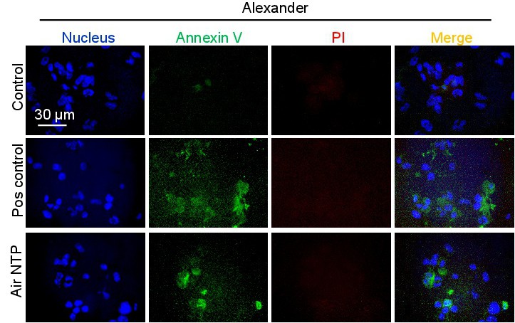
Fig. 9. Alexander cells were treated with NTP for 60 s and then 6 h after treatment cells were labelled with Hoechst nuclear stain - blue dye, annexin V - green dye and propidium iodide - red dye. Labelled cells were imaged with fluorescence microscopy. Representative images out of three independent experiments are shown. Positive control - 2 µM staurosporine for 4 h.